
AFM in Life Sciences: Molecular Characterization
Seeing and Measuring Biological Structures at the Nanoscale
Download the 2026 Molecular Characterization AFM whitepaper (PDF)
Table of Contents
1. Introduction
2. Why Use AFM to Characterize Biological Molecules?
3. Applications of AFM for Imaging Molecules
3.1. DNA Structure and Quality
3.2. Protein Folding and Aggregation
3.3. Gene Therapy Vectors
4. AFM Solutions for Characterizing Molecules
1. Introduction
Atomic force microscopy (AFM) gives life science researchers a practical way to visualize and measure individual biological molecules—including DNA, proteins, and gene therapy vectors—with quantitative, three-dimensional nanoscale detail.
Unlike transmission and scanning electron microscopy (TEM/SEM), which generally require high vacuum and often heavy-metal staining, AFM can examine biological structures without those requirements. And, while light microscopy remains diffraction- limited, AFM can measure surface topography and mechanical properties at the nanoscale in liquids.
2. Why Use AFM to Characterize Biological Molecules?
AFM provides quantitative, sub-nanometer three-dimensional topography and mechanical data in air or liquid. For the examples in this white paper, dry-sample preparation takes less than 30 minutes, sample volumes are typically 1–5 µL, no heavy-metal contrast agents are required, and consumable costs are under $30 per run. The data presented here were acquired using AFMWorkshop AFMs costing less than $70,000. These AFMs have a very low annual operating cost that depends primarily on operator probe consumption. AFMWorkshop systems do not require maintenance contracts and are designed to be robust and intuitive, allowing customers to perform any routine maintenance independently. Optional remote support and maintenance contracts are available. AFMWorkshop also offers affordable system refurbishment and upgrade packages.
3. Applications of AFM for Imaging Molecules
3.1. DNA Structure and Quality
Characterizing DNA is important for quality control in synthetic biology and genevtherapy. Gel electrophoresis can indicate fragment length, conformational changes, and relative quantities, but results are typically presented as bands or smears. AFM adds a direct view of individual DNA molecules, helping researchers examine construct cleanliness, fragmentation, supercoiling, and folding. The M13 examples shown here illustrate how AFM can reveal structural differences that are not visible from a band pattern alone.
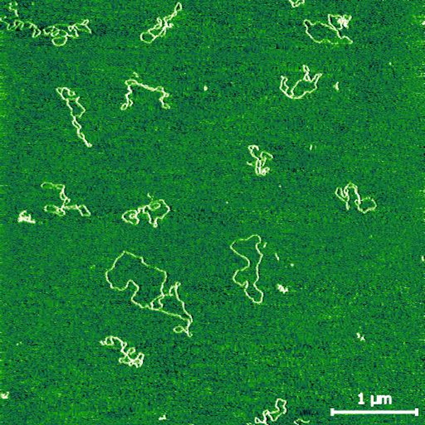
M13 plasmid supercoiling and fragmentation (dsDNA, 4.8 kbp)
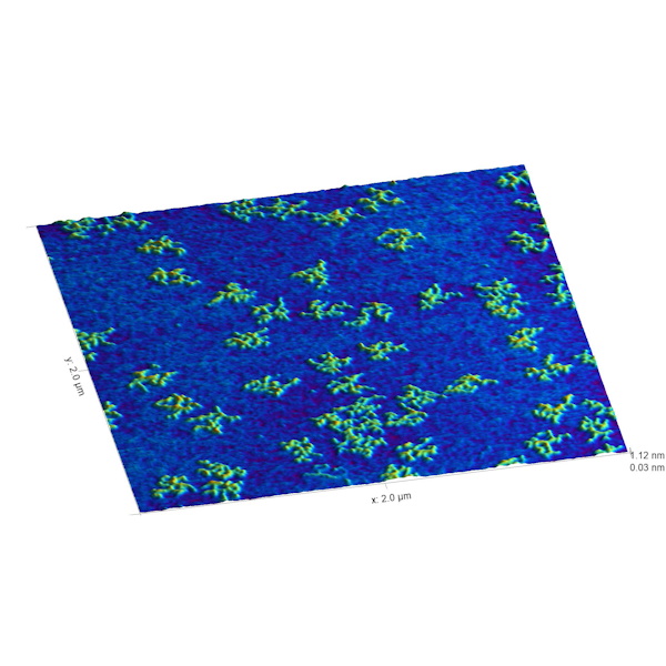
3D representation of folded M13 ssDNA (4.8 kb)
3.2. Protein Folding and Aggregation
Proteins and protein complexes play central roles in laboratory assays and biological processes. AFM is particularly useful for examining protein folding and aggregation. Proteins can be deposited onto mica and imaged to assess monomer morphology, aggregate structure, and aggregate size. The PrP amyloid examples below show fibril aggregation, image-based segmentation of individual aggregates, and the resulting size distribution.
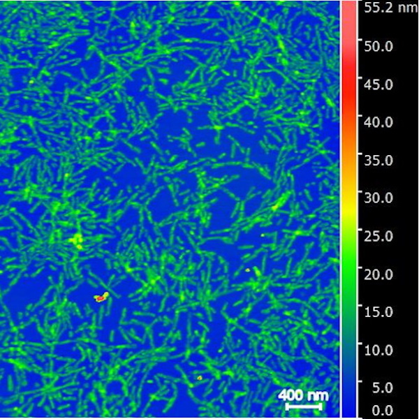
PrP fibril aggregation
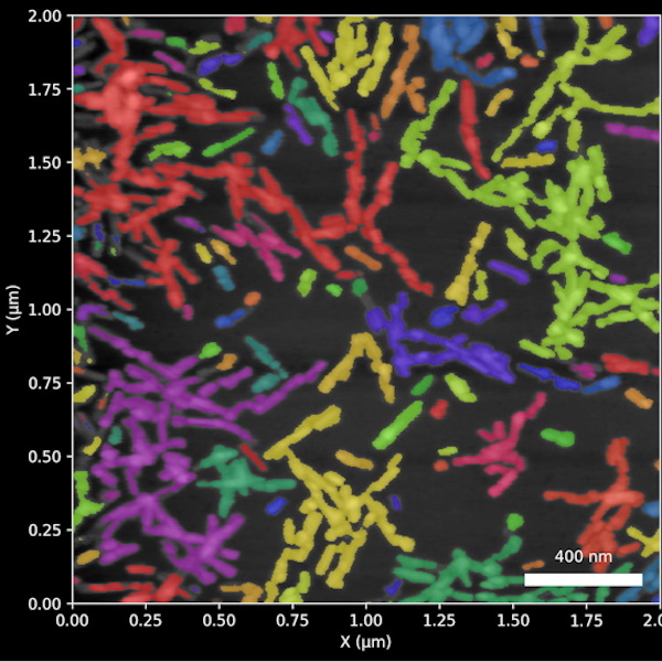
PrP aggregate segmentation
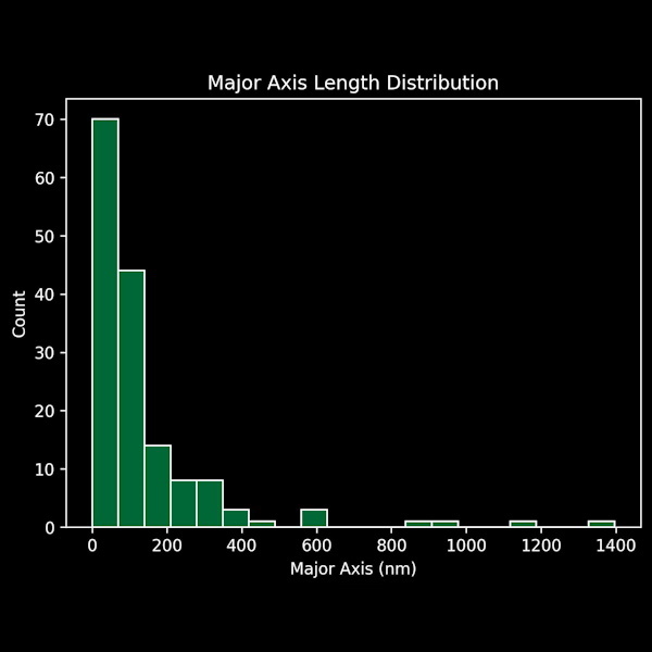
PrP aggregate major axis length
3.3. Gene Therapy Vectors
AFM characterization of DNA and proteins comes together in the analysis of gene therapy vectors. Adeno-associated virus (AAV) vectors are approximately 26 nm in diameter and package a single-stranded DNA (ssDNA) genome of about 4.8 kb. AFM measurements can be used to assess both vector and genome integrity. In unmodified samples, measurements can show the relative frequency of free ssDNA and viral particles, along with particle-height distributions. With straightforward sample-preparation protocols, free ssDNA can also be distinguished from capsid-bound DNA.
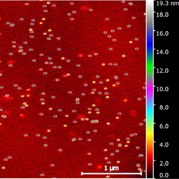
Height indexed AAV particles with released DNA (red “clouds”)
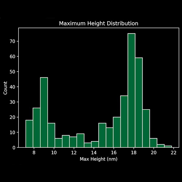
AAV height distribution
4. AFM Solutions for Characterizing Molecules
AFM is increasingly used to characterize molecular populations in research settings, including biochemistry and molecular biology, and in industrial quality control for applications such as gene therapy and biosensors. For biosensor manufacturing in particular, AFM offers a non-destructive approach: sensor substrates can be removed from the production line, qualified, and returned to production in less than an hour. AFMWorkshop systems are designed to integrate into laboratory and production workflows at a comparatively accessible cost, without requiring prior AFM expertise.
AFM makes it possible to move beyond indirect molecular measurements and examine biological structures directly. By bringing DNA, protein, and vector characterization into one accessible nanoscale imaging workflow, it offers researchers and manufacturers another practical tool for evaluating molecular structure and sample integrity.
AFMWorkshop offers high-performance AFMs that are robust, intuitive to use, and affordable for a wide range of laboratories. Our innovative AFM designs emphasize the features most important for scanning biological materials, without the added complexity and cost of unnecessary features often found in higher-priced systems.
The images presented in this white paper were measured by AFMWorkshop customers primarily using the HR AFM. The HR 2D AFM is a great benchtop AFM for measuring dry molecular structures.
HR AFM
The HR AFM includes features such as rapid approach, a universal probe holder, a top- view optical microscope, and an off-axis optical microscope. Pricing for the stage and the scanner starts at $49,976.
HR-2D AFM
The compact HR-2D AFM features a noise floor of less than 0.03 nm while maintaining a small footprint suitable for space-constrained laboratories. Pricing for the stage and a scanner starts at $42,215.

The HR AFM includes the AFM Stage, control electronics, and software for high resolution scanning.
AFMWorkshop offers several vibration solutions, and installation packages.
